Bioimaging Facility
The Leduc Bioimaging Facility provides equipment and training dedicated to high-resolution imaging in the life sciences.
Bioimaging Facility
The Leduc Bioimaging Facility provides equipment and training dedicated to high-resolution imaging in the life sciences.
About

The Leduc Bioimaging Facility, open to all investigators, provides equipment and training dedicated to high-resolution imaging in the life sciences. The facility includes a Cryo Transmission Electron Microscope, a Scanning Electron Microscope, four Fluorescence Microscopes, two automated Slide Scanners, four Confocal Laser Scanning Microscopes, a Multiphoton Microscope, a spinning disk High-Content Screening System, and software for image analysis. The facility also maintains equipment for sample preparation, including a critical point dryer, sputter coater, and microtomes for ultrathin sectioning.
The Bioimaging Facility is a Service Center in the Division of Biology and Medicine and provides microscopes and supporting equipment in Sidney Frank Hall for Life Sciences and the Laboratories for Molecular Medicine.
Learn more about the history of the facility below.
The Bioimaging Facility is listed within CoresRI.org, a searchable directory of core facilities in Rhode Island.
Services and Instruments
Microscopes in Sidney Frank Hall for Life Sciences
185 Meeting Street: Rooms 105, 106, 107, 109, 111, 113, 115, 117
BMC: Rooms 005
Microscopes in Laboratories for Molecular Medicine
70 Ship Street: Rooms 111, 133
Service Request and Reservations
Reserve a microscope using the iLab calendars
If you already have an ILabs account, click the link below to create an equipment reservation.
Rates
FY26 Rates
| Service | Units | Internal Academic Rates* | External Academic Rates |
|---|---|---|---|
| Confocal | |||
| Tundra Cryo (First 4 Hours) | Hours | $64 | $101 |
| Tundra Cryo (Additional Hours) | Hours | $20 | $31 |
| Olympus FV3000 Confocal Microscope | Hours | $52 | $82 |
| Zeiss LSM 800 Confocal | Hours | $51 | $81 |
| Zeiss LSM 880 Confocal (LMM) | Hours | $51 | $82 |
| Multiphoton | |||
| Olympus FV-1000-MPE Multiphoton Microscope | Hours | $62 | $99 |
| Fluorescence | |||
| Nikon Ti2-E High-Content Analysis Fluorescence Microscope (LMM) | Hours | $31 | $49 |
| Nikon Ti2-E Widefield Fluorescence Microscope (SFH) | Hours | $28 | $44 |
| Zeiss Axiovert 200M Fluorescence Microscope (SFH) | Hours | $28 | $44 |
| Electron Microscopy | |||
| Apreo VS SEM - (First 4 Hours) | Hours | $64 | $102 |
| Apreo VS SEM - (Additional Hours) | Hours | $18 | $29 |
| Slide Scanner | |||
| Olympus VS200 Slide Scanner (LMM) | Hours | $40 | $63 |
| Olympus VS200 Slide Scanner (SFH) | Hours | $40 | $64 |
| High Content | |||
| Notice: Opera Phenix New User and New Uses Incentive Program. Click here to apply | |||
| Opera Phenix High-Content Screening System | Hours | $72 | $114 |
| Sample Prep | |||
| Ultra-microtome | Hours | $40 | $64 |
| Critical Point Dryer | Hours | $18 | $29 |
| Sputter Coater | Hours | $21 | $34 |
| Diamond Knife | Hours | $23 | $37 |
| Pelco Biowave | Hours | $115 | $184 |
| Technical Assistance | |||
| Technical Assistance | Hours | $45 | |
| Training | Session | $283 | $452 |
*Rates for Brown and Rhode Island Academic and Hospital Affiliates
Effective 11/18/2024
Contacts/Location
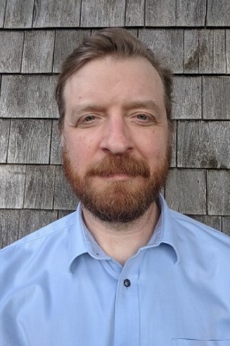
Microscopist/Facility Manager
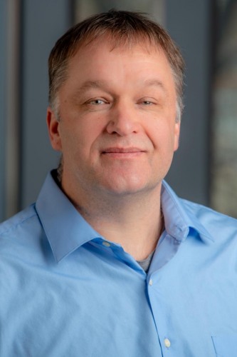
Facility Director
The Bioimaging Facility maintains microscopes at the following two locations
The Bioimaging Facility at Sidney Frank Hall
Room 106, 107, 109, 111, 113, 115, 117
185 Meeting Street
Providence, RI 02912
The Bioimaging Facility at the Laboratories for Molecular Medicine
Room 111, 133
70 Ship Street
Providence, RI 02903
Access & Training
The facility largely operates on a self-service model: students, staff and faculty are trained on a specific microscope and can then use the microscope independently. Full-service assistance in microscopy is available. The facility provides training in microscopy, image analysis, and ultrathin sectioning. If you'd like to use one of the microscopes in the facility, you must first schedule training, before signing up on the microscope.
To schedule a training or request assistance in imaging please contact Geoff Williams.
To request assistance in sample preparation (e.g. fixation, mounting, sectioning, staining, immunolabeling) and imaging, please contact the Molecular Pathology Core.
Molecular Pathology Core
The Molecular Pathology Core provides all-inclusive histology and histopathology services, equipment and training. The core houses an automated tissue processor, a paraffin embedding center, automated microtomes, a cryostat, a vibratome and a multiheaded light microscope.
Resources for Grants
Acknowledgment
This facility was supported in part by grants from the National Institutes of Health (NIH), Brown University's Division of Biology and Medicine and Provost's office.
* Please acknowledge NIH support. E.g. in publications that made use of the SEM:
The Thermo Apreo VS SEM was purchased with a high-end instrumentation grant from the Office of the Director at the National Institutes of Health (S10OD023461).
The Zeiss 710 Laser Scanning Confocal Microscope was purchased with a high-end instrumentation grant from the Office of the Director at the National Institutes of Health (S10RR023693).
The Tundra Cryo-TEM was purchased with a high-end instrumentation grant from the Office of the Director at the National Institutes of Health (S10OD032301).


