Division of Biology and Medicine
BioMed Core Facilities
Nikon Ti2-E Fluorescence Microscope for High-content Imaging
High-content imaging system, based on an inverted microscope and equipped for imaging standard slides, culture dishes, and microplates (e.g. 96-well plates).
Nikon Ti2-E Fluorescence Microscope for High-content Imaging
High-content imaging system, based on an inverted microscope and equipped for imaging standard slides, culture dishes, and microplates (e.g. 96-well plates).
Overview
Laboratories for Molecular Medicine, Room 111
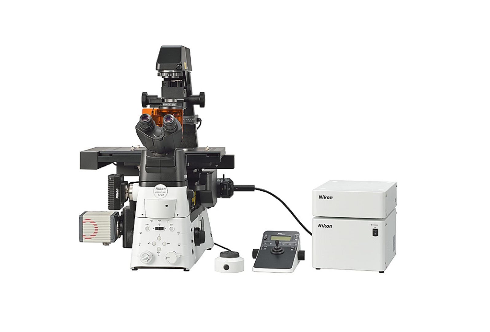 The Nikon Ti2-E fluorescence microscope is a high-content imaging system, based on an inverted microscope and equipped for imaging standard slides, culture dishes, and microplates (e.g. 96-well plates). The microscope includes 'Perfect Focus' for autofocus applications, a Photometrics Prime 95B sCMOS camera for imaging fluorescence, a 16 megapixel Nikon DS-Ri2 color camera for imaging histology slides, a motorized stage and NIS-Elements High Content (HC) software for multiwell plate acquisition.
Operation
Objectives:
- 4x Plan Apochromat Lambda, N.A. 0.2, W.D. 20 mm, F.O.V. 25 mm
- 10x Plan Fluor Phase Contrast DL, N.A. 0.3, W.D. 16 mm, F.O.V. 25 mm
- 20x Super Plan Fluor ELWD, N.A. 0.45, W.D. 6.9-8.2 mm, F.O.V. 22 mm, DIC, Correction Collar 0-2 mm
- 40x Super Plan Fluor ELWD, N.A. 0.6, W.D. 2.8-3.6 mm, F.O.V. 22 mm, DIC, Correction Collar 0-2 mm
- 60x Oil Plan Apochromat Lambda Phase Contrast DM, N.A. 1.4, W.D. 0.13 mm, F.O.V. 25 mm, Ph3
Resources
Nikon
Reservations
Microscopes in the Bioimaging Facility can be reserved using iLab calendars. All facility users are invited to use the system, which requires a one-time registration.