The facility has been expanded significantly in recent years: the electron microscopes were upgraded for digital imaging and additional imaging systems were purchased, including three fluorescence microscopes, three confocal laser scanning microscopes, and a multiphoton microscope. Training and equipment are currently offered at two locations: in Sidney Frank Hall for Life Sciences and the Laboratories for Molecular Medicine. The facility maintains nine main imaging systems and serves more than 250 users.
Elizabeth H. Leduc, PhD
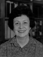
Elizabeth H. Leduc, a biologist who greatly advanced our understanding of cellular structure and function using electron microscopy, who is known to most people as “Dukie,” spent her childhood near the Canadian border in Vermont, and graduated with a Bachelor of Science degree from the University of Vermont in 1943. Subsequently, she obtained a Master’s degree from Wellesley College and a Ph.D. from Brown. She held an NIH postdoctoral fellowship at Brown from 1948-49, taught Anatomy at Harvard Medical School from 1949-1953, and then returned to Brown where she spent the remainder of her career. And what an illustrious career that was: a Brown Alumni Magazine writer described her as being at “…the forefront of three fields. An effective teacher, a successful administrator, and a scientist with international credentials.”
Fortunately, these abilities were recognized. In 1964, she was appointed as a full Professor of Biology – the third woman to attain that rank in the almost two hundred years of Brown’s existence, and the first in Biology. She was appointed Frank L. Day Professor of Biology in 1975.
From 1973-1977, she served as a Dean of Biological Sciences. Reflecting on this, she commented, “Originally, I had hoped to do my share of the administrative work of the medical school closer to my retirement, but I was persuaded otherwise by Dr. Galletti’s view that no administrator should have to give up teaching or research.” Demanding as this range of activities was, Dukie was successful in them all: the Division of Biology and Medicine continued to expand during her deanship (at a time when the University itself was in poor financial shape and was undergoing budget cuts), she co-taught a large course in Histology and she continued her research. Towards the end of her tenure as Dean, she was appointed by President Gerald Ford to the President’s Committee on Science and Technology – the only woman on the nine-person committee. This committee reviewed the entire structure of federal science, a task which Dukie herself described as “a mountainous job.”
Dukie had broad interests in cell biology. Her bibliography begins with a series of papers co-authored with her Ph.D. mentor, J. Walter Wilson. The papers focus on mitosis in the liver and deal with the effects of different stimuli on mitotic activity, and the production of polyploid nuclei and multinucleate cells in the liver. This interest in the cell biology of the liver was maintained throughout her career so that during the 1970’s she was researching liver pathology caused by the oxidation products of cholesterol while also experimenting with new chemotherapeutic agents, which inhibited mitotic activity of hepatomas.
While her research on the liver was ongoing, Dukie was also pioneering an important new methodology, cytochemistry. In 1951 and 1952, she co-authored papers describing the use of histochemistry to localize acid and alkaline phosphatase. Later, she and Wilhelm Bernhard pioneered the use of water-soluble embedding media and ultrathin frozen sections.
Dukie’s collaboration with Bernhard was part of a wider collaboration with investigators at the Institut de Reserches Scientifiques sur le Cancer at Villejuif, France. Dukie described this institute as being like “a little scientific NATO” and relished the two months that she spent there during most summers from 1959 to the mid 1980’s. Her fluency in French led to another responsibility at Brown – she examined Ph.D. candidates in Biology to determine their competency in French. With her collaborators in France, she produced a series of papers on the formation of perichromatin granules and the effects of quinacrine on nuclear structure. In 1984, shortly before her retirement, she brought together many of her scientific interests in a paper entitled “Immunocytochemical identification of nuclear structures containing snRNPs in isolated rat liver cells.”
During her academic career, Dukie was a widely known and influential scientist. She was a member of 12 professional societies, served on several editorial boards, and was a member of the National Advisory General Medical Sciences Council of the NIH. From 1972-1976, she was a consultant for the National Cancer Institute of Canada. Of special interest to MCB graduate students, she served on the Council of the American Society for Cell Biology from 1976-1980 and during her career she was mentor for 7 Ph.D. candidates. Many of you will use the Leduc Electron Microscopy suite, named in her honor.
Dukie was a gracious person who traveled widely, and enjoyed good company and fine food. In 1967, a reporter asked whether all her research on the liver had affected her taste for this organ: she said no, “I love it. There’s a little restaurant in Paris ----”
For all her achievements, Dukie was an approachable person who delighted in showing schoolchildren around her mouse colony, and who during the mid 1980’s served as a Randall Counselor for sophomores and an Associate Dean of the College. She continued to interact with students, staff and faculty long into retirement.
Elizabeth H. Leduc, 88, passed away on January 30th, 2010 and is dearly missed by all who knew her.
We thank Peter Heywood for Dukie’s biography.