Evident Scientific FV-1000-MPE Multiphoton Microscope
Deep optical sectioning of fluorescently labeled samples with SIM photoactivation
Evident Scientific FV-1000-MPE Multiphoton Microscope
Deep optical sectioning of fluorescently labeled samples with SIM photoactivation
Overview
Sidney Frank Hall, Room 106B
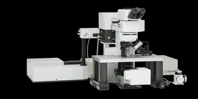
In April 2013, the Bioimaging Facility set up an Olympus FV1000-MPE multiphoton laser scanning microscope (also referred to as a 'two-photon' or '2P' microscope). This microscope is designed for high-resolution imaging of fluorescence at depths that are inaccessible to standard fluorescence or confocal microscopes. The multiphoton microscope excites fluorescent indicators solely in the plane of focus and stacks of images can be acquired for 3D reconstruction and visualization. The multiphoton microscope is based on an Olympus BX61wi upright microscope and is equipped with a Mai Tai HP tunable laser (690-1020 nm). A SIM scan unit is available for simultaneous photostimulation with a 405 nm laser. In addition, 458, 488, 515, 559, and 635 nm laser lines are available for sequential photoactivation and imaging. The microscope is equipped with four non-descanned detectors (2 PMTs and 2 GaAsP detectors), an encoded Prior Z deck with a scanning stage, and 10x (NA 0.30, WD 3.3 mm), 25x (NA 1.05, WD 2 mm), and 60x (NA 1.00, WD 2 mm) objectives. An analog input box with mapping and multipoint software is available for combined imaging and electrophysiology experiments.
As of August 2017 we have added a special Second Harmonics Filter cube that allows direct visualization of unstained Collagen fibers either isolated (in tendons or digested) or w/in tissue (cardiac for example). The filter is a 405/40 allowing selection of a stimulation wavelength between ~780 and ~840 nm.
Operation
Reservations
Reserve a microscope using the iLab calendars
Reserve a microscope using the iLab calendars