Division of Biology and Medicine
BioMed Core Facilities
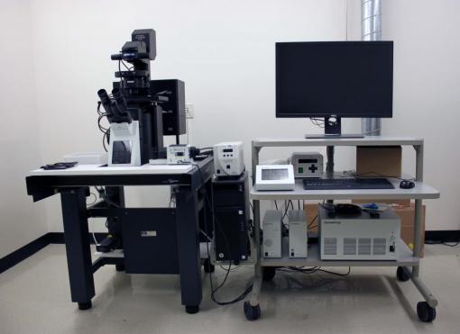
Evident Scientific FV3000 Confocal Microscope
Optical sectioning of samples labeled with fluorescent indicators. Equipped with a resonant scanner for fast optical sectioning
Evident Scientific FV3000 Confocal Microscope
Optical sectioning of samples labeled with fluorescent indicators. Equipped with a resonant scanner for fast optical sectioning
Overview
Sidney Frank Hall, room 111
 This confocal microscope can acquire thin optical slices of fluorescent samples. It has an inverted microscope configuration and is equipped with a resonant scanner for fast optical sectioning and 4 detectors, two standard Metal Alkalide and two high sensitivity GaAsP detetors.
- LASER DIODE 405NM,CW 50MW
- LASER DIODE 488NM,CW 20MW
- LASER DIODE 561NM,CW 20MW
- LASER DIODE 640NM,CW 40MW
Operation
Objectives on the FV3000RS:
| Mag | Correction | NA | HFW @ 1x | WD (mm) | Immersion Medium |
|---|---|---|---|---|---|
| 1.25x | UPlan Apochromat | 0.04 | 10.18 mm | 5.0 | Air/Dry |
| 10x | UPlan Super Apochromat | 0.4 | 1.27 mm | 3.1 | Air/Dry |
| 20x | UPlan Super Apochromat | 0.75 | 0.600 mm | 0.60 | Air/Dry |
| 30xS | UPlan Super Apochromat | 1.05 | 0.424 mm | 0.80 | Silicone Oil |
| 60xS2 | UPlan Super Apochromat | 1.3 | 0.212 mm | 0.30 | Silicone Oil |
Reservations
Reserve a microscope using the iLab calendars
Reserve a microscope using the iLab calendars