Image Analysis SFH
Software for image analysis and 3D visualization
Image Analysis SFH
Software for image analysis and 3D visualization
Overview
Sidney Frank Hall, Room 106C
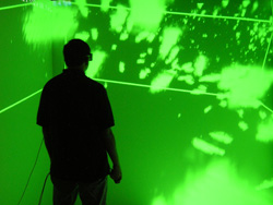
FluidVis. Collaborations between Brown's Computer Science Department, the Center for Computation and Visualization (CCV) and the basic science departments in the Division of Biology and Medicine have led to the development of the research software CaveVOX, a fully-immersive interactive three-dimensional visualization application that runs in the Brown Cave at 180 George Street. CaveVOX has been in use for several years and helped understand some 3D stacks more quickly and led to discoveries overlooked with desktop visualization tools. FluidVis is a new software that provides a semi-immersive experience similar to CaveVOX and is available for use now in the Bioimaging Facility. FluidVis runs on hardware acquired through an OVPR seed grant awarded to Professor Kristi Wharton. Both CaveVOX and FluidVis create an intuitive virtual environment for analyzing stacks of images in 3D and are ideally suited for the analysis of samples with considerable 3D complexity. For example, networks of neural connections can be difficult to analyze in two dimensions, but stand out bright and clear in a 3D environment. A natural user interface makes changing viewpoints and exploring or tuning visualization settings easy and fast.
For a demo or training on FluidVis, please contact Robbert Creton by phone at 401-863-9646 or by email at: Robbert_Creton@brown.edu. Please feel free to bring a stack of images for the training. The images should be in TIF format, one series per folder, with sequentially numbered names. For additional information about CaveVOX and 3D visualization systems at the Center for Computation and Visualization, please visit the CCV website at http://www.ccv.brown.edu/ . For more information about FluidVis, please visit http://www.fluiditysoftware.com/.
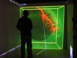
Investigator uses CaveVOX to explore 3D datasets acquired by confocal microscopy
Sidney Frank Hall Room 106B also houses two workstations for image analysis:
1) Metamorph. The first workstation contains MetaMorph software (version 7.0), which can be used for deconvolution (digital removal of out-of-focus light), 3-D reconstruction, brightness measurements, cell counting, colocalization analysis, fluorescence / brightfield overlay, FRET analysis, morphometry, motion analysis, particle tracking, and timelapse measurements.
2) Confocal Software. The second workstation contains image analysis software for the Zeiss Confocal Microscope (LSM Imager) and Leica Confocal Microscope (LCS) that can be used for image viewing, export of TIF images, fluorescence / brightfield overlays, quantitative measurements, colocalization analysis, and 3D reconstruction.