XROMM Service
XROMM Service
Overview
The W.M. Keck Foundation XROMM Facility biplanar room at Brown University consists of two Varian model G-1086 x-ray tubes, two EMD Technologies model EPS 45-80 pulsed x-ray generators, two Dunlee model TH9447QXH590 image intensifiers (16" diameter), and two Phantom v10 high-speed digital video cameras. The x-ray tubes are suspended from the ceiling by tube cranes and the IIs are mounted on mobile gantries. The components are set up such that the two x-ray beams intersect each other close to the IIs. A treadmill, trackway or other animal support/enclosure is placed such that the research animal performs the behavior of interest (running, jumping, flying) in the volume where the x-ray beams intersect.
The Brown University system can deliver pulsed x-ray generation at up to 100 Hz, and can record in continuous x-ray generation mode at up to 1000 fps. With the Phantom v10 cameras, the pixel resolution is 1800x1800. Overall resolution of the imaging chain is about 2 line pairs/mm. Radio-opaque beads can be tracked to within ±0.1 mm in 3D space. The x-ray system was designed and integrated by Marty Kulis of Imaging Systems and Service, Painesville OH (mkulis@issi-na.com; 440-724-8002).
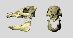 aces from CT, laser scanning, or MRI. Each bone is an object that can be manipulated individually in computer animation space. These models are specific to each individual study subject (human or animal).
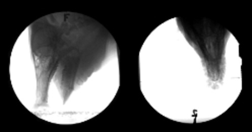 o high-speed, biplanar x-ray movies. In some studies it is possible to surgically implant radiopaque beads into the bones for
 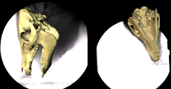 The data on bone shape and bone movement are combined into an XROMM animation. Rigid body kinematics from the x-ray movies are applied to the bone models to re-animate the actual movement that was performed by the individual subject at the time of recording.
Operation
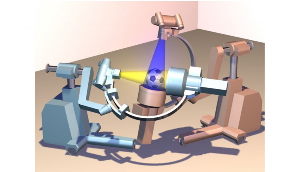
Diagram of mobile C-arm fluoroscopes, retrofitted for high-speed imaging. New 30-cm image intensifiers have been installed and the original cameras have been replaced with high-speed video cameras. A standard international (size 5) soccer ball is included for scale. Radiological Imaging Services, Hamburg PA (800-748-2040) has experience refurbishing and retrofitting C-arms for high-speed imaging.
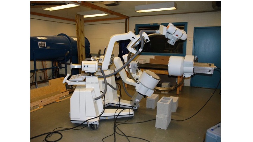
Photograph of mobile C-arm fluoroscopes, retrofitted for high-speed imaging. New 30-cm image intensifiers have been installed and the original cameras have been replaced with high-speed video cameras. Radiological Imaging Services, Hamburg PA (800-748-2040) has experience refurbishing and retrofitting C-arms for high-speed imaging.
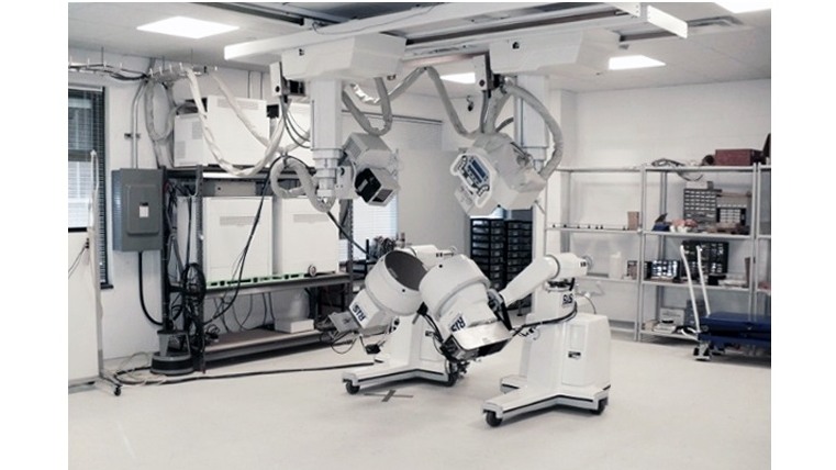
The W.M. Keck Foundation XROMM Facility at Brown University. Two x-ray tubes are suspended from the ceiling by tube cranes and two image intensifiers are mounted on mobile gantries. Independent movement of the four components gives the system a lot of flexibility in physical configuration. Here the system is configured to collect two oblique, ventro-dorsal views.
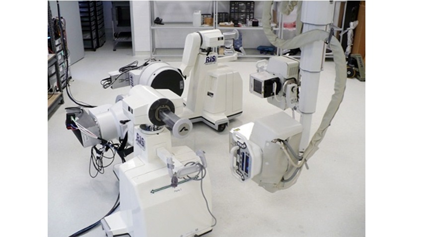
The W.M. Keck Foundation XROMM Facility at Brown University. Independent movement of the four components gives the system a lot of flexibility in physical configuration. Here the system is configured to collect two oblique, medio-lateral views.